Resp. Éric Sibert
X-rays provide a valuable tool for analysis as they have a deep penetration in the light materials like water or polymer membranes. This gives the opportunity to investigate electrochemical systems in situ i.e. with electrolyte and under electrochemical potential. By using high brilliance X-ray sources provided by third generation synchrotrons, one can probe metal surfaces. In this framework, we are performing in situ surface X-ray diffraction at the surface of single crystal electrodes, at ESRF, on beamlines CRG-D2AM and ID03, in collaboration with Institut Néel (Yvonne Soldo and Maurizio De Santis) and Jakub Drnec from ESRF, resp.
We developed two spectro-electrochemical cells. The first one is a thin layer cell that is optimized for low electrolyte contribution and that can be used with sample booth horizontal and vertical. The second one is a reverse hanging meniscus configuration that provides a better electro-chemical configuration, especially for kinetic investigations.
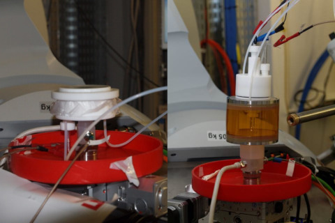
Figure 1: Electrochemical cells for surface X-ray diffraction. Left: thin film. Right: inverted hanging meniscus.
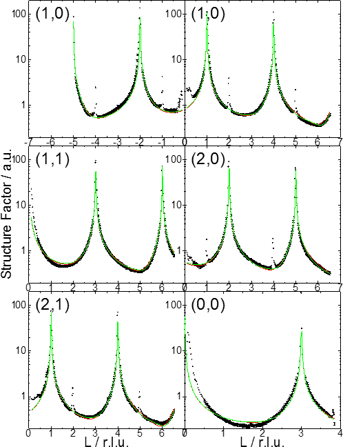
Figure 2 : CTRs of the Ir(111) surface at 0.6 VRHE in 0.05 M H2SO4. Black: measurements. Green: modelling. Measurements on beamline ID03/ESRF.
Former work on the monitoring of hydrogen insertion in Pd nanofilms on Pt(111) : doi:10.1016/j.electacta.2013.06.095
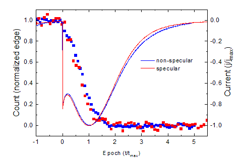
Figure 3: Two simultaneous measurements of the electrochemical deposition current of 1 ML Pd on Pt(111) (solid lines) and the diffraction signal (points) in anti-Bragg positions.
On figure 3, time scales are normalized to the maximum deposition current. Non-specular signal is related to ordered deposit while specular is sensitive to ordered and non-ordered deposits.
Growth mechanisms of Pd nanofilms electrodeposited onto Au(111): an in situ grazing incidence X-ray diffraction study : doi:10.1039/c5cp05985b
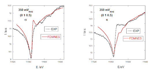
Figure 4: SRXRD spectra of Pt(111) in 0.1 M H2SO4 at +350 mVRHE. Parallel (left) and perpendicular (right) polarizations.
X-rays provide a valuable tool for analysis as they have a deep penetration in the light materials like water or polymer membranes. This gives the opportunity to investigate electrochemical systems in situ i.e. with electrolyte and under electrochemical potential. By using high brilliance X-ray sources provided by third generation synchrotrons, one can probe metal surfaces. In this framework, we are performing in situ surface X-ray diffraction at the surface of single crystal electrodes, at ESRF, on beamlines CRG-D2AM and ID03, in collaboration with Institut Néel (Yvonne Soldo and Maurizio De Santis) and Jakub Drnec from ESRF, resp.
We developed two spectro-electrochemical cells. The first one is a thin layer cell that is optimized for low electrolyte contribution and that can be used with sample booth horizontal and vertical. The second one is a reverse hanging meniscus configuration that provides a better electro-chemical configuration, especially for kinetic investigations.
Figure 1: Electrochemical cells for surface X-ray diffraction. Left: thin film. Right: inverted hanging meniscus.
Surface structure
The determination of a surface structure, at a given electrochemical potential, is performed by measuring several crystal truncation rods (CTRs) of a sample. Then, the data modeling with the ROD software allows to determine the organization of the surface (nature and occupancy of the atomic layers, interatomic distances, disorder...). The following figure shows the rods of an Ir(111) electrode. Figure 2 : CTRs of the Ir(111) surface at 0.6 VRHE in 0.05 M H2SO4. Black: measurements. Green: modelling. Measurements on beamline ID03/ESRF.
Former work on the monitoring of hydrogen insertion in Pd nanofilms on Pt(111) : doi:10.1016/j.electacta.2013.06.095
Kinetic measurements
The goal is to follow in situ and in real time modifications at the electrode surface. As surface transformations are much faster than CTRs measurements duration, we can only record one or two points in reciprocal space during a kinetic measurement. We choose points with high sensitivity to surface structure, usually in anti-Bragg position i.e. in the middle of two Bragg peaks, along a CTR. We did a first campaign on beamline CRG-D2AM for the deposition and the dissolution of Pd on Pt(111). Figure 3: Two simultaneous measurements of the electrochemical deposition current of 1 ML Pd on Pt(111) (solid lines) and the diffraction signal (points) in anti-Bragg positions.
On figure 3, time scales are normalized to the maximum deposition current. Non-specular signal is related to ordered deposit while specular is sensitive to ordered and non-ordered deposits.
Growth mechanisms of Pd nanofilms electrodeposited onto Au(111): an in situ grazing incidence X-ray diffraction study : doi:10.1039/c5cp05985b
Surface Resonant X-Ray Diffraction
SRXRD couples surface X-Ray diffraction, widely used to solve the atomic structure at the surface of single crystals, to X-Ray absorption near edge spectroscopy (XANES), highly sensitive to the oxidation states. The experimental approach is supported by the theoretical ab initio calculations of the FDMNES software developped by Yves Joly (Institut Néel), recently implemented with the simulation of surface diffraction experiments and now under development for electrochemical interfaces description. We did first measurements on beamline CRG-D2AM for the Pt(111) surface in sulfuric acid solution. Figure 4: SRXRD spectra of Pt(111) in 0.1 M H2SO4 at +350 mVRHE. Parallel (left) and perpendicular (right) polarizations.